Scientific Program

Day 1 :

Biography:

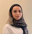
Nirvana Khalaf Mansour is an endodontic specialist and graduated from Cairo University in 2009 with a bachelor degree. She earned her master degree in endodontic 2016 and doctorate  in 2021.She was worked for five years in suez military hospital 2014-2019. And works as Endodontist private practice in Cairo area Egypt and founder of dr.nirvana dental clinic. Currently member of scientific committee of princess Fatma Academy and lecture in ministry of health, Egypt.

Abstract:

Vital pulp therapy has been known as one of the treatment options to preserve pulp vitality after being exposed by trauma or caries. Aim: This experiment explored the effect of injectable-Platelet Rich Fibrin on marginal adaptation of two pulp capping agents (Mineral Trioxide Aggregate and Bioactive Bone Graft). A total of 64 teeth were used out of 8 healthy male beagle dogs. The teeth were randomly assigned into four groups, they were exposed and capped with different capping agents. Group A; capped with Mineral Trioxide Aggregate (MTA), Group B; capped with MTA+ i-PRF, Group C; capped with Bioactive Bone Graft (BBG), Group D; capped with BBG+i-PRF. Finally the access cavity was restored with Intermediate Restorative Material (IRM). At each predetermined interval, the dogs were sacrificed (1 month, and 3 months). The samples were then prepared for electron microscopic scanning evaluation. To compare between the gap percentage of four groups at each interval, Kruskal-wallis test; was used. Mann-Whitney U test; was used to pair-wise comparison when Kruskal-wallis test is significant. Bonferroni’s correction was utilized for the pair-wise comparisons. Statistical significance was considered at P < .05. The data revealed that after one and three months the best values were recorded in groups B (MTA+ i-PRF) and D (BBG+ i-PRF), in relation to the lowest gap area between the capping materials and dentin, followed by group C (BBG), with the least value recorded in group A (MTA). The findings from the current study suggested that i-PRF provided a better marginal adaptation of either MTA or BBG to the pulp and dentin, which improved with time from one month to three months

Keynote Forum

Mohammad A. AlShammari,

Kuwait Ministry of Health, Kuwait

Keynote: Why are we still saving teeth? The revival of microsurgery in the implant epoch

Time : 9:00:00

Biography:

Mohammad A. AlShammari is an Endodontist recently graduated and qualified from the Kuwait Board of Endodontics. He qualified from Jordan University of Science & Technology in 2016, then he worked at Kuwait Ministry of Health during his training year he applied for Kuwait board program & got accepted. During the first of the program, he was awarded the MFD (RCSI) from the Royal College of Surgeons in Ireland in 2018, Elected as the Chief Resident. Alshammari has a special interest in endodontic microsurgery and had treated many varieties of cases during the past few years.

Abstract:

In the dental implant epoch, the trend seems to be to extract compromised teeth and replace them with implants. Yet, the long-term prognosis of teeth might not be comparable with the prognosis of dental implants. Complications, failures, and diseases such as peri-implantitis are not rare, and, despite the belief, implants are not 99% successful. Other treatment options that aim to save compromised teeth such as microsurgery, intentional replantation, and autotransplantation should be considered on an individual basis. With the advent of surgical techniques and instruments, the clinical and radiographic outcomes of endodontic surgery have been dramatically improved, these treatments have competing success rates to dental implants. And more importantly, it retains the natural tooth in the dentition for a longer period of time. 

  • Dentistry and Dental Health
Location: Dubai

Chair

Nirvana Khalaf Mansour

Dubai

  • Oral and Maxillofacial Surgery
Location: Dubai

Chair

Mohammad A. AlShammari,

Kuwait Ministry of Health, Kuwait

Session Introduction

Reema Sharaf

Prince Sultan Military Medical City, Saudi Arabia Saudi Arabia

Title: Complete dental and oral rehabilitation and management of patient after Laryngeal cancer treatment
Speaker
Biography:

Reema Sharaf has completed her BDS in 1997 – 1998 King Saud University in Riyadh KSA. AGD USC 2002, Saudi Board in Advanced Restorative Dentistry 2009, Consultant in Restorative and Esthetic Dentistry till the present, Deputy of Restorative Dentistry Department from 2016-2019.a

Abstract:

Head and neck cancer is a term used to describe a number of different malignant tumors that develop in or around the throat, larynx, nose, sinuses, and mouth. Laryngeal cancer is cancer of the larynx, which is a part of the throat.

Successful management of laryngeal cancer depends on careful pretreatment evaluation of the patient, and  appropriate treatment selection for patients with this disease.

Management of the head and neck cancer patients requires a multidisciplinary team. Treatment  includes radiation, chemotherapy, and surgery.  Patients with malignant disease need careful dental treatment planning prior to radiation therapy  and/or chemotherapy. In this presentation, we  report a case of a patient with laryngeal cancer, who did not have a chance to follow dental treatment recommendations for both pre- and post radiation dental management. The aim is to create awareness for the clinician to evaluating the patient for the disease related issues that may affect the oral cavity and dentition, and to know what treatment options do we have in this case. The treatment option used in this case for oral rehabilitation and improvement of his quality of life.

Speaker
Biography:

Aween Auda Ablahad is a dentist and oral and maxillofacial pathology specialist, graduated from University of Mosul in 2009 with a bachelor degree. She earned her master degree in oral and maxillofacial pathology from college of dentistry/Hawler Medical University in 2016 and now she is a PhD candidate in the same specialty, she has her own private dental clinic since 2013.  She is a lecturer and a member in college of dentistry, University of Duhok, Iraq.

Abstract:

Introduction: Implication of human papilloma virus (HPV) in the carcinogenesis of oral squamous cell carcinoma (OSCC) is debatable subject, p16 overexpression indicates active HPV infection in oropharyngeal squamous cell carcinoma (OPSCC) but in OSCC such relation still needs to be studied. Therefore, we aimed to evaluate the frequency of HPV in OSCC patients in the capital of Kurdistan region of Iraq and its concordance with p16 overexpression. We retrieved Eighty-six formalin-fixed paraffin-embedded (FFPE) samples of OSCC from multi large pathological centers that located in the capital of Kurdistan, we utilized immunohistochemistry (IHC) to detect the HPV by anti-HPV high risk antibody correlated it with p16 overexpression, besides, twenty FFPE samples of healthy gingiva were used as control in this study. Chi square and Fisher’s exact tests were used for correlating the HPV status and p16 overexpression with clinicopathogical patient’s data. The concordance between HPV and p16 overexpression was evaluated by kappa agreement and spearman rank correlation. The frequency of HPV in OSCC patients were 15.1%, tongue was the most common site affected by HPV infection, other patient data including age, gender, grade and stage did not show significant correlation with neither anti-HPV nor P16 antibodies. The concordance level between p16 overexpression and the HPV status according to kappa agreement was (κ = 0.221, p= 0.034), Moreover, the correlation according to spearman correlation coefficient was (r = 0.229, p= 0.034), with 46.15% sensitivity and 80.82% specificity. We concluded that HPV infection is still low in Erbil and p16 biomarker has only diminutive significance as a predictor of HPV infection in the OSCC patients.

  • Dental Surgery
Location: Dubai

Chair

Mohammad A. AlShammari

Kuwait Ministry of Health, Kuwait

Session Introduction

Samar Jambi

Ministry of Health, Saudi Arabia

Title: Partial enucleation of the Nasopalatine canal for implant placement
Speaker
Biography:

Samar Jambi is a certified consultant in prosthodontics and a Board Certified consultant of implant dentistry practicing at the North Jeddah Centre of Dental Specialties, Jeddah, Saudi Arabia. She earned her Bachelor degree in Dentistry from King Abdulaziz University, Saudi Arabia in 1995, Master degree in Restorative dentistry from Leeds University, UK in 2002, PhD degree in Fixed Prosthodontics from Leeds University, UK in 2008. She did her Fellowship in Implant Dentisrty King Abdulaziz University, Saudi Arabia in 2015. Samar Jambi is a member of the Saudi Dental Society as well.

Abstract:

Statement of the Problem: One of the anatomical obstacles for dental implants placement in the upper jaw is the nasopalatine canal. The neurovascular contents include nasopalatine nerve, terminal branch of nasopalatine artery and anastomoses with greater palatine nerve and artery. This gives innervation and vascularization for the upper anterior region from the right canine to the left canine. Unfortunately, after extraction of the anterior maxillary teeth, high resorption rate happens on the area. In addition, presence of the incisive canal jeopardizes the ideal position of the implants placement. As a result, enucleation of the canal content and then replaced by bone graft or substitute is necessary to improve the bone bed. Neurological impairment of the soft tissue such as paresthesia or dysesthia may exist after enucleation of the canal. In order to prevent any change in sensation and having bone augmentation at the same time, partial removal of the canal content (coronal 2/3 of its contents) and placement of allograft bone is presented in this case report. Re-entry surgery after six month of the surgical site showed an adequate bone. An implant was placed then a prosthetic restoration was placed. After one year of follow up the implant was successful and no sensory disturbances were shown. A comparison was presented before and after the treatment of the case. Clinical photos and CT figures were included.  

Day 2 :